


2Diagnostiskt Centrum Hud Stockholm, Apelbergsgatan, Sweden.
3Diagnostiskt Centrum Hud Malmo, Hjalmaregatan Sweden
4Department of Oncology-Pathology, Karolinska Institutet, Karolinska University Hospital, Sweden
Objective: To characterize CMM patients diagnosed at two dermatology clinics, to calculate and compare lead times for patients found to have CMM with those with benign nevi and to calculate the melanoma detection rate.
Methods: A retrospective, cross-sectional study at two dermatology clinics in Sweden. All patients diagnosed with CMM at these clinics were included; 102 in Stockholm and 22 in Malmo. A control group consisting of patients with benign pigmented lesions were age and gender matched with the CMM group. Data were collected from medical records.
Results: Patients with CMM had shorter lead times than control patients from first visit to excision (median 5 vs 17 days, p = 0.003) and from first visit to histopathological diagnosis (27.5 vs 40 days, p = 0.003). Females waited longer than males from referral/booking to first visit (25 vs 15.5 days, p = 0.014). The CMM detection rate was 1.4 %.
Conclusions: Our results imply that the dermatologist can shorten lead times when a CMM is suspected. The CMM detection rate was higher than reported from Euro melanoma weeks. Together this indicates that a health care system with easy access to dermatologists can be effective. The CMM patients had shorter waiting times than nationally, suggesting that general improvements in Sweden are possible.
Key words: melanoma; early diagnosis; waiting times; secondary prevention
A patient who seeks medical attention for a pigmented skin lesion is, in current practice in most parts of Sweden, primarily managed by the Primary Care Physicians (PCPs). The PCPs are, in turn, expected to refer the patient to specialist care in case of high clinical suspicion of skin cancer [7]. Studies have, however, shown that dermatologists are better at diagnosing pigmented skin lesions than PCPs regarding diagnostic accuracy, lead times and costs [7,8]. The difficulties in diagnosing skin lesions in primary care often result in surgical excision of benign naevi, which increases the health care costs [9]. In Sweden, the yearly direct costs of removing benign skin lesions amount to approximately 300 million SEK (36.7 million USD). The costs for treatment of CMM are estimated to 166 million SEK (20.3 million USD) per year [9].
In addition to being cost-beneficial, the better diagnostic accuracy among dermatologists may contribute to shorter time to diagnosis and treatment of CMM, and thereby result in reduced morbidity and mortality. A Swedish study [10] found that CMM patients primarily examined by dermatologists had shorter lead times to primary excision and histopathological diagnosis, compared to patients first consulting a PCP. A US retrospective single-centre study showed that patients having an established contact with a dermatologist had thinner CMM or CMM in situ, than those irregularly visiting a dermatologist [11].
The median lead time in Sweden from first doctor's examination to diagnosis of CMM is 37 days [12]. Swedish recommendations state that 80% of patients should receive a definite diagnosis within 28 days from their first medical visit [13]. Data from 2010-2013 show that this was due for only 40% of CMM patients [13].
In 2012 in Stockholm, and two years later in Skane, the so-called Health Choice Dermatology ("VardvalHud") was introduced in Sweden. This model allows patients to turn directly to a dermatology clinic bypassing primary care, and enabled the start of Diagnostiskt Centrum Hud (DCH). DCH comprise of two open-care dermatology clinics, one located in Stockholm and one in Malmo. Due to the positive association between early detection and patient prognosis, together with the tendency described above of higher efficiency when patients can access dermatology specialists directly, we aimed to explore lead times and detection rates for CMM patients in this new model, "Health Choice Dermatology".
|
Event |
A |
Referral or booking [1] |
B |
First visit at clinic |
C |
Primary excision |
D |
Preliminary histopathological report |
E |
Final histopathological report |
F |
Referral for wide excision |
G |
Wide excision at clinic |
When comparing patient and tumour characteristics between the three study groups, Chi-square test for categorical data (or Fischer's exact test when categories contained less than 5 subjects) and Mann-Whitney U test for continuous data were performed. Patient and tumour characteristics were likewise tested for potential associations with age (divided in patients ≤ 60 and > 60 years), gender and presence or absence of referral from other health services. The Mann Whitney U test was used to compare differences in lead times as well as to test for potential association between patient and tumour characteristics, including gender, age, and presence or absence of referral from a PCP. The significance level was set to p < 0.05.
To calculate the melanoma detection rate, the number of diagnosed CMM at the clinic in Stockholm between March 2012 and August 2014 were divided with the total number of individual consecutive patients undergoing dermatoscopy during the same period. This was done using the monthly reports sent to the Stockholm County Council from DCH, containing all billable medical services
|
Stockholm |
Malmo |
|
|
|
|
Melanoma
|
Control |
Melanoma |
p-value1 |
p-value2 |
Total no. of patients |
102 |
204 |
22 |
|
|
Males, n (%) |
53 (52) |
106 (52) |
10 (46) |
1.0003 |
0.5803 |
Age, y |
|
|
|
|
|
Mean (SD) |
60.0 (16.7) |
59.4 (16.9) |
61.5 (18.4) |
0.9274 |
0.5344 |
Range |
23-90 |
23-93 |
24-88 |
|
|
Contact with clinic, n (%) |
|
|
|
<0.0013 |
0.1243 |
Referral |
55 (54) |
61 (30) |
8 (36) |
|
|
Self-appointment |
46 (45) |
143 (70) |
14 (64) |
|
|
Not available |
1 (1) |
|
|
|
|
Anatomical site, n (%) |
|
|
|
<0.0013 |
0.6645 |
Trunk |
45 (44) |
67 (33) |
11 (50) |
|
|
Head and neck |
26 (26) |
28 (14) |
3 (14) |
|
|
Lower extremities |
17 (17) |
18 (9) |
4 (18) |
|
|
Upper extremities |
14 (14) |
2 (1) |
4 (18) |
|
|
Multiple sites |
|
69 (34) |
|
|
|
Not available |
|
20 (10) |
|
|
|
Melanoma subtype [6], n (%) |
|
NA |
|
|
0.1675 |
SSM |
69 (68) |
|
10 (46) |
|
|
LM |
19 (19) |
|
8 (37) |
|
|
LMM |
5 (5) |
|
1 (5) |
|
|
Other |
9 (9) |
|
3 (14) |
|
|
Invasive, n (%) |
55 (54) |
NA |
12 (55) |
|
0.9273 |
T-categories |
|
NA |
|
|
0.2865 |
Tis |
47 (46) |
|
10 (46) |
|
|
T1 |
41 (40) |
|
7 (32) |
|
|
T2 |
8 (8) |
|
2 (9) |
|
|
T3 |
5 (5) |
|
2 (9) |
|
|
T4 |
0 |
|
1 (5) |
|
|
Breslow thickness[7], mm |
|
NA |
|
|
0.2534 |
Mean (SD) |
0.89 (0.69) |
|
1.52 (1.38) |
|
|
Median (range) |
0.70 (0.10-3.40) |
|
0.83 (0.30-4.15) |
|
|
Clark, n (%) |
|
NA |
|
|
0.4465 |
I |
47 (46) |
|
10 (46) |
|
|
II |
17 (17) |
|
4 (18) |
|
|
III |
24 (24) |
|
4 (18) |
|
|
IV |
13 (13) |
|
3 (14) |
|
|
V |
0 |
|
1 (5) |
|
|
Ulceration, n (%) |
5 (5) |
NA |
0 |
|
0.5845 |
CMM patients referred to the clinic in Stockholm were slightly older than those who were self-referred, mean age 61.5 years vs 57.0 years (p = 0.142). Corresponding figures for the control group were 63.5 years vs 57.5 years
(p = 0.018).
Patients with CMM in Malmo had a significantly shorter waiting time than CMM patients in Stockholm for the period between preliminary (D) and final histopathological report (E). Furthermore, patients in Malmo waited shorter time from final histopathological report (E) to wide excision at the clinic (G).
In the CMM group in Stockholm, women waited longer than men from the date of referral or booking (A) to first visit at the clinic (B) (median 25 versus 15.5 days, p = 0.014). For the same lead time, patients older than 60 years waited longer than young patients. (26.5 vs 15 days, p = 0.027).Patients referred to the clinic waited longer than self-referred patients (27 versus 14 days, p=0.002). Patients with thick CMM (> 1.00 mm, n = 13) had significantly shorter lead times in Stockholm when compared to patients with thin CMM. The difference in lead times was significant between first visit (B) and primary excision (C) (median 0 versus 7.5 days, p = 0.009), excision (C) and preliminary histopathological report (D) (6 versus 16 days, p = 0.008) and between first visit (B) and final histopathological report (E) (13 versus 33 days, p = 0.001).
|
Melanoma, Stockholm |
Control, Stockholm |
Melanoma, Malmo |
|
|
|||
Interval |
Mean |
Median (range) |
Mean |
Median (range) |
Mean |
Median (range) |
p-value1 |
p-value2 |
A à B |
26.5 |
20.0 (0-192) |
22.3 |
21.0 (0-97) |
23.9 |
21.5 (8-46) |
0.588 |
0.678 |
B à C |
18.1 |
5.0 (0-153) |
28.9 |
17.0 (0-242) |
11.7 |
6.5 (0-55) |
0.003 |
0.881 |
C à D |
15.0 |
15.5 (3-34) |
20.0 |
19.0 (6-34) |
16.7 |
15.0 (5-45) |
<0.001 |
0.539 |
D à E |
5.0 |
3.5 (0-39) |
0.8 |
0.0 (0-14) |
2.7 |
0.0 (0-29) |
<0.001 |
0.006 |
E à F |
9.5 |
8.0 (-10-40) |
NA3 |
4.0 |
4.0 (1-9) |
|
0.131 |
|
E à G |
30.5 |
24.0 (3-91) |
NA3 |
18.9 |
19.0 (7-36) |
|
0.050 |
|
B à E |
38.3 |
27.5 (4-166) |
51.4 |
40.0 (11-263) |
31.1 |
27.5 (6-71) |
0.003 |
0.772 |
A à F |
74.1 |
59.0 (5-366) |
NA |
46.0 |
46.0 (33-59) |
|
0.499 |
|
A à G |
95.6 |
84.5 (27-231) |
NA |
67.8 |
65.5 (56-84) |
|
0.180 |
|
Patients in Malmo waited a significantly shorter time than patients in Stockholm between preliminary and final histopathological report. The reasons for this may relate to differences between the two cities in regards to histopathological workload or working processes. The difference in lead time between final histopathological report and wide excision, where patients in Malmo waited fewer days in median, approached statistical significance. There were no differences in patient and tumour characteristics between the clinics explaining the differences in waiting times. Considering the relatively small cohort in Malmo, due to the shorter study period there, our results should be analysed with caution, but indicate that lead times, especially regarding the histopathology review, could potentially be shortened in Stockholm.
Women with CMM waited a longer time than men in Stockholm from referral or booking to first visit at the clinic. This is interesting considering a study by Hajdarevic et al. [10], reporting that women waited shorter times than men. In that study, however, the case mix was different from ours, with women having significantly more in situ tumours and thinner invasive CMM [10]. For the same lead time, we also found that CMM patients > 60 years waited longer than younger patients in Stockholm. According to the Swedish NBHW, older people with cancer, including CMM, wait longer for examination and treatment than younger people [14]. This inequality needs to be addressed, as there is an association between age and tumour thickness, with thicker tumours especially seen among older men living alone [15]. Thus, early detection is of outmost importance also among older patients.
The observation that referred patients, compared to the selfreferred, in both groups in Stockholm had longer lead times from first contact to first visit could be influenced by the handling of self-referrals at the clinic. A handful of self-referred patients were scheduled for a visit the same day, due to last-minute cancellations from other patients. There were also a few days, e.g. at the Melanoma Week in May, when the clinic was open for drop-in appointments for patients with pigmented skin lesions.
For year 2013, the Swedish NBHW reported that the waiting time for melanoma patients from first visit to definitive diagnosis was in median 38 days in Stockholm and 28 days in Skane [12]. In our study, the median waiting time from first visit to final histopathological diagnosis was 27.5 days in both Stockholm and Malmo. However, there was a difference between the national report and our study in how the date of the definitive diagnosis was defined. The national report used the date when the patient received information about their diagnosis [12]. In our study, such information was often missing in the medical records. We therefore used the date of the final histopathological report as the date of diagnosis. In reality, the time until the patient received information about the definitive diagnosis is longer. The Swedish NBHW states that 80 % of patients should receive a diagnosis within four weeks from their first doctor's visit [13]. In our study, 54% of the CMM patients in Stockholm and 50% of the patients in Malmo had a waiting time of less than four weeks for the interval between first visit and final histopathological report. These results do not fulfil the recommendations, but still exceed the all-country data from 2010-2013, where 40 % of patients met the criteria, with reservation for the mentioned divergence in definitions of lead times [13].
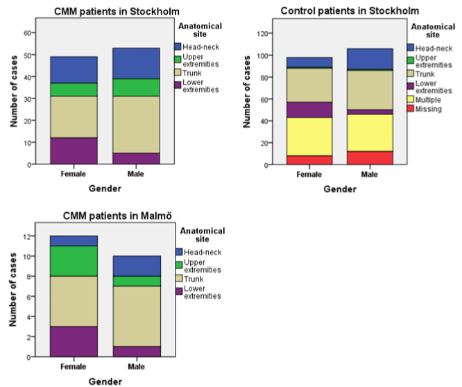
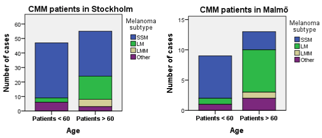
The characteristics of the CMM's found were in concordance with published data. Tumours on the trunk were more common among men, and tumours on the lower extremities more common among women. Similar results have been presented in quality reports for Sweden since the early 90s and the differences are thought to correspond to the sunbathing and clothing habits for each sex [16]. Patients > 60 years had more lentigo subtypes and more CMM in the head- and neck region, when compared to younger patients. Since lentigo subtypes usually appear in regions chronically exposed to sunlight, i.e. the head and neck, these two findings are linked to one another [17]. Additionally, because of the slow growth pattern of LM and LMM, these subtypes are more common among older patients [17].The majority of the CMM diagnosed at the two clinics were in situ or thin tumours (T1). This finding is encouraging as identifying CMM at an early stage minimizes the risk of local recurrence and metastasis [4].
An unexpected finding was that about 20% of patients in both CMM groups underwent subtotal incisional biopsies before or instead of primary excision. This treatment option is not recommended in guidelines as previous studies have shown an increased risk of incorrect staging in the histopathological analysis [16, 18]. Our results exceed that in another Swedish study, where 10% of patients underwent biopsies [10], but can be explained and motivated by the fact that most biopsied CMM were of lentigo subtypes.
A detection rate of 1.4% might be considered fairly low. With the potential benefit in having high access to dermatologists not being limited to identifying the more obvious CMM, but also to find the more obscure, early cases of thin CMM or even in situ cases, there is obviously still a need to excide a high number of benign lesions in order not to miss a CMM. As mentioned above, there are indications of dermatologists being more effective than PCPs [10] and, when consulting a dermatologist, the efficiency is higher if the patient regularly sees the same dermatologist [11]. In regions with low access to dermatologists, tele-dermatoscopy could help to triage the patients that really needed to be examined by a specialist [20].
- Tzellos T, Kyrgidis A, Mocellin S, Chan AW, Pilati P, Apalla Z. Interventions for melanoma in situ, including lentigo maligna. Cochrane Database Syst Rev. 2014;12:CD010308. doi: 10.1002/14651858. CD010308.pub2.
- Cancer incidence in Sweden 2012: new cancer cases diagnosed in 2012. Stockholm : The National Board ; 2014. Available from: http://www.socialstyrelsen.se/Lists/Artikelkatalog/Attachments/ 19291/2013-12-17.pdf
- Causes of Death 2013. Stockholm : The National Board ; 2014. Available from: http://www.socialstyrelsen.se/Lists/Artikelkatalog/ Attachments/19736/2015-2-42.pdf
- Balch CM, Gershenwald JE, Soong SJ, Thompson JF, Atkins MB, Byrd DR , et al. Final version of 2009 AJCC melanoma staging and classification. J Clin Oncol. 2009;27(36):6199-6206. doi: 10.1200/JCO.2009.23.4799. Epub 2009 Nov 16.
- Bordeaux JS, Lu KQ, Cooper KD. Melanoma: prevention and early detection. Semin Oncol. 2007;34(6):460-466.
- Krige JE, Isaacs S, Hudson DA, King HS, Strover RM, Johnson CA. Delay in the diagnosis of cutaneous malignant melanoma. A prospective study in 250 patients. Cancer. 1991;68(9):2064-2068.
- Chen SC, Pennie ML, Kolm P, Erin M Warshaw, Eric L Weisberg, Katherine M Brown, et al. Diagnosing and managing cutaneous pigmented lesions: primary care physicians versus dermatologists. J Gen Intern Med. 2006;21(7):678-682. doi: 10.1111/j.1525- 1497.2006.00462.
- Mc Kenna DB, Marioni JC, Lee RJ, Prescott RJ, Doherty VR. A comparison of dermatologists’, surgeons’ and general practitioners’ surgical management of cutaneous melanoma. Br J Dermatol. 2004;151(3):636-644.
- Lindelof B, Hedblad MA, Ringborg U. [Nevus or malignant melanoma? Correct diagnostic competence results in lower costs]. Lakartidningen. 2008;105(39):2666-2669.
- Hajdarevic S, Hornsten A, Sundbom E, Isaksson U, Schmitt-Egenolf M. Health-care delay in malignant melanoma: various pathways to diagnosis and treatment. Dermatol Res Pract. 2014;2014:294287.
- Cheng MY, Moreau JF, Mc Guire ST, Ho J, Ferris LK. Melanoma depth in patients with an established dermatologist. J Am Acad Dermatol. 2014;70(5):841-846. doi: 10.1016/j.jaad.2013.10.060. Epub 2014 Mar 12.
- Waiting times in cancer care: report December 2014. Stockholm : The National Board ; 2014. Available from: http://www.socialstyrelsen. se/Lists/Artikelkatalog/Attachments/19616/2014-12-11.pdf
- Regional Cancer Centre in Collaboration . National quality report of diagnosis 2010-2013 - From the National hudmelanomregistret [ Internet] . Cancer Center ; 2014 [ cited 2015 03 10]. Available from: http://www.cancercentrum.se/globalassets/cancerdiagnoser/hud/ kvalitetsregister/20140929_melanom_rapport_2010-2013.pdf
- National Board . Waiting times in cancer care - from referral to start of treatment [ Internet] . National Board ; 2011. [ cited 2015 05 28] . Available from: http://www.socialstyrelsen.se/Lists/Artikelkatalog/ Attachments/18872/2012-10-28.pdf
- Eriksson H, Lyth J, Mansson-Brahme E, Margareta Frohm-Nilsson, Christian Ingvar, Christer Lindholm , et al. Later stage at diagnosis and worse survival in cutaneous malignant melanoma among men living alone: a nationwide population-based study from Sweden. J Clin Oncol. 2014;32(13):1356-1364.
- Regional Cancer Centre in Collaboration . Malignant melanoma - the national program of care [ Internet] . Cancer Center ; 2014 [ cited 2015 03 09]. Available from : http://www.cancercentrum.se/globalassets/ cancerdiagnoser/hud/vardprogram/natvp_malignt_melanom_ rev.2015-01-19lang.pdf
- Cummins DL, Cummins JM, Pantle H, Silverman MA, Leonard AL, Chanmugam A. Cutaneous malignant melanoma. Mayo Clin Proc. 2006;81(4):500-507.
- Karimipour DJ, Schwartz JL, Wang TS, Bichakjian CK, Orringer JS, King AL, et al. Microstaging accuracy after subtotal incisional biopsy of cutaneous melanoma. J Am Acad Dermatol. 2005;52(5):798-802.
- van der Leest RJ, de Vries E, Bulliard JL, Paoli J, Peris K, Stratigos AJ, et al. The Euromelanoma skin cancer prevention campaign in Europe: characteristics and results of 2009 and 2010. J Eur Acad Dermatol Venereol. 2011;25(12):1455-1465. doi: 10.1111/j.1468- 3083.2011.04228.x. Epub 2011 Sep 23.
- Borve A, Dahlen Gyllencreutz J, Terstappen K, Johansson Backman E, Aldenbratt A, Danielsson M, et al. Smartphone teledermoscopy referrals: a novel process for improved triage of skin cancer patients. Acta Derm Venereol. 2015;95(2):186-190. doi: 10.2340/00015555- 1906.

